


Panoramic X-rays are one of the most important diagnostic imaging procedures in dentistry. They are widely used as they can show the entire dentition, the jawbone, and surrounding anatomical structures on a single two-dimensional image.
Its frequent use is mainly due to the fact that it provides very quick, easily understandable information about the entire oral cavity, and with modern X-ray machines, the radiation dose is negligible.
Because many people get scared when going to the dentist Panoramic X-ray Many users don't know what an application is or why it's needed, nor do they understand how it's made or what it entails. Therefore, we've decided to put together a professional article to explain this topic.
We felt it was important to share our own experiences and knowledge, as well as doing so in plain language, so below everyone is guaranteed to find answers to their questions.
What is a panoramic X-ray?
A panoramic X-ray is a so-called extraoral, which means an X-ray taken from outside the oral cavity. The device has precise operation, and the patient simply has to stand still while the machine slowly scans the oral cavity from all sides.
This is important because a panoramic X-ray provides a complete picture not just of a single tooth or region, but of the entire dentition, the jawbone, and the associated joints, which is essential for certain dental procedures.
What does a panoramic X-ray show?
A panoramic X-ray can provide comprehensive diagnostic information from a dental perspective about the entire oral cavity. For example, it shows:
- the entire dental arch and the position of the teeth, as well as any missing teeth;
- significant cavities, damage, the condition of root-treated teeth, and any inflammations;
- the condition of the jaw, including the jawbones and associated bone structure;
- possible malformations, malformations of the teeth and traumatic fractures.
For most dental problems, panoramic X-rays are therefore sufficient for diagnosis and treatment, but in complicated cases 3D CBCT is needed to more accurately assess the three-dimensional anatomy, for example.
The panoramic X-ray process step-by-step
In most dental practices – where such a device is available – panoramic X-rays are considered a routine examination. The process generally unfolds as follows:
Preparations:
As with all X-ray examinations, it is important that the patient removes any surrounding jewellery, such as piercings, earrings and necklaces in the face or mouth, and is then given a special protective gown to protect them from radiation.
Positioning
For a panoramic X-ray, it is essential that the dentition is visible in the correct plane. For this reason, the patient must place his head in a jaw and forehead rest, and the technician often uses a Plexiglas support to stabilise the front teeth.
Recording:
The patient must remain still while the X-ray is being taken. The machine usually takes 15-20 seconds to take a panoramic image.
Result
As most modern dentists now use digital X-rays, the image is immediately displayed on the screen, which the dentist can evaluate and save for later use.
When do you need a panoramic X-ray?
Panoramic X-rays are usually necessary before the following dental complaints or procedures:
First dental check-up:
Especially during the first dental examination, it may be necessary to examine the entire oral cavity, even in the absence of complaints, in order to document the anatomical background of the dentition and jaw.
Status check:
In the follow-up and condition check for certain dental procedures, such as a root canal treatment, X-rays can be a very useful aid.
Tooth decay
If the decay is extensive, involving several teeth, and a physical examination is not possible to decide on the appropriate intervention, a panoramic X-ray can help to make a diagnosis.
Inflammations:
Because X-rays can detect inflammation, cysts and nodules, they have important diagnostic value in determining the location and extent of suspected inflammation.
Periodontal disease:
Severe periodontal disease is most often accompanied by bone loss. The extent of this can be easily revealed with the help of a panoramic X-ray.
Surgical intervention:
Before oral surgery procedures (for examplewisdom tooth extraction(in any case) it can always be important to know the condition and position of the affected teeth, thus avoiding possible complications.
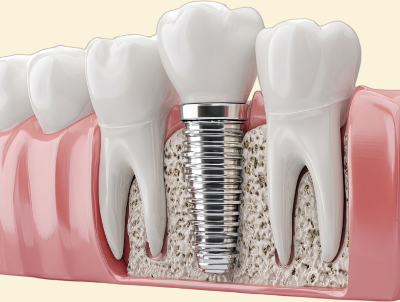
Implantological planning:
Although 3D CB CT scans are often required prior to implantation, panoramic X-rays may be sufficient for a preliminary bone structure assessment or when planning the need for bone replacement.
Tooth developmental abnormalities:
In development disorders of the teeth, such as wisdom teeth not growing properly or the absence of a tooth germ, X-rays can help to better understand the problem before any potential interventions.
Orthodontics:
To plan orthodontic treatment, it is essential to know the parts of the teeth that are not visible. Panoramic X-rays are particularly helpful in this respect, as they can present the entire oral cavity to the dentist.
When is a panoramic X-ray not sufficient?
As it turns out, panoramic X-rays are an effective diagnostic tool in many cases, however, there are instances where they are not sufficient and 3D CBCT is required. Let's look at some examples of this:
- Spatial information is needed about the teeth and their surroundings, for example when an implant is inserted or the exact location of the nerve canal.
- If very small details, such as a root crack, need to be explored.
- If a more precise anatomical exploration is required, for example for a complex surgical procedure.
Panoramic X-ray or 3D CBCT?
As the 3D has been mentioned several times aboveCBCTconcept, it's worth being aware of the difference between it and panoramic X-rays!
It is important to note that there are cases and interventions where both imaging modalities may be needed to ensure the precision of the intervention, but there are still fundamental differences between the two methods.
- Whereas a panoramic X-ray is typically a quick and inexpensive solution, which can provide a 2D image of the teeth with a low radiation dose,
- while CBCT can reconstruct the dentition in 3D, giving dentists a much more detailed anatomical picture, and is therefore considered a more expensive procedure than X-rays.
Advantages of panoramic X-ray
A panoramic X-ray is a very useful imaging method for detecting certain dental abnormalities. Its advantages include:
Fast:
The test requires no special preparation, and the complete scan is usually completed in a few minutes.
Painless:
As an imaging method, it is a non-invasive procedure, completely free from pain and other discomforts.
Comprehensive
It allows you to see the entire dentition, jaws and joints in a single scan.
Digital:
In modern dental practices, panoramic X-rays operate on a digital principle, meaning the image can be viewed, saved, and transmitted immediately.
Safe
Modern X-ray machines have been developed to work with very low radiation exposure, so the examination is completely safe.
Cost-effective
In terms of value for money, panoramic X-rays are considered the best diagnostic tool in dentistry.
The radiance of panoramic X-rays
As with all X-ray diagnostics, radiation dose is an important issue in panoramic X-rays.
We have already pointed out several times that modern digital X-ray machines work with very small amounts of so-called ionizing X-rays.
For panoramic X-rays, for example, the average dose is around 10-25 µSv, which is roughly the same as a day or two of natural background radiation for comparison.
We are exposed to continuous radiation in our everyday lives, which amounts to about 2500-3000 µSv per year in Hungary.
Compared to this, the radiation emitted by panoramic X-rays is negligible, but also insignificant compared to a head and neck CT scan, where the average radiation dose can reach 2000 µSv.
It is also important that modern X-ray machines always work with automated exposure settings, taking into account, for example, gender and age, and always use the lowest possible minimum value for imaging.
Frequently asked questions
What Is the Difference Between a Panoramic X-ray and a Traditional Dental X-ray?
While conventional dental X-rays (intraoral X-rays) are only suitable for exploring a small area, one or two teeth, panoramic X-rays can provide a high-resolution, comprehensive image of the entire dental arch.
Can panoramic X-rays be used during pregnancy?
During pregnancy, all imaging tests except ultrasound are prohibited. Exceptions can only be made in cases of strong justification, in the case of urgent intervention, by using the lowest possible radiation dose or extra radiation protection.
How often can a panoramic X-ray be taken?
There is no general rule for this; each case is assessed based on individual risk. Since digital X-ray images can be saved and retrieved at any time, repeating them in the short term is unnecessary.
When should I have a 3D CBCT instead of a panoramic X-ray?
When spatial information and more detail is needed, such as when an implant is being placed or for a more complex surgical procedure.
What can distort a panoramic X-ray image?
Distortion may be caused primarily by patient movement and improper head positioning, and less commonly by the proximity of metal objects or equipment malfunction.
What preparations are required for a panoramic X-ray?
This imaging procedure does not require significant preparation, but it is advisable to attend the examination without necklaces, earrings, and piercings. This is because removing them – along with glasses – is important for safe and good quality imaging.
What is the difference between modern digital panoramic X-rays and the old analogue methods?
The biggest difference is that digital panoramic X-ray is not only faster, but also works at a lower dose rate, so it captures a safer and even better quality image that is immediately visible on the connected monitor, so it can be used immediately.
Can the panoramic X-ray also show nodules and cysts?
For larger lesions, yes, but for small or not clearly identifiable nodules and cysts, further investigation is needed.
How much does a panoramic X-ray cost?
The price of the panoramic X-ray in the NaturaDent dental office is 15 000 HUF.